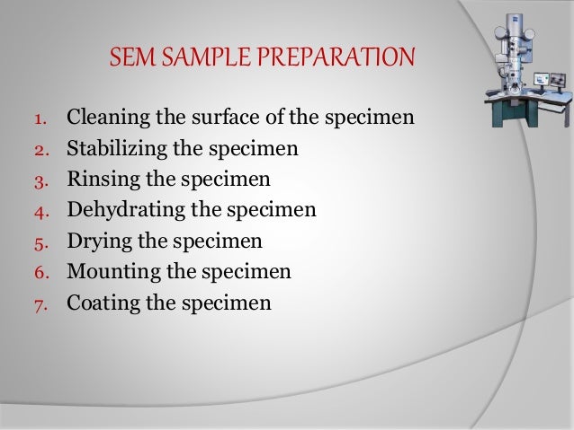
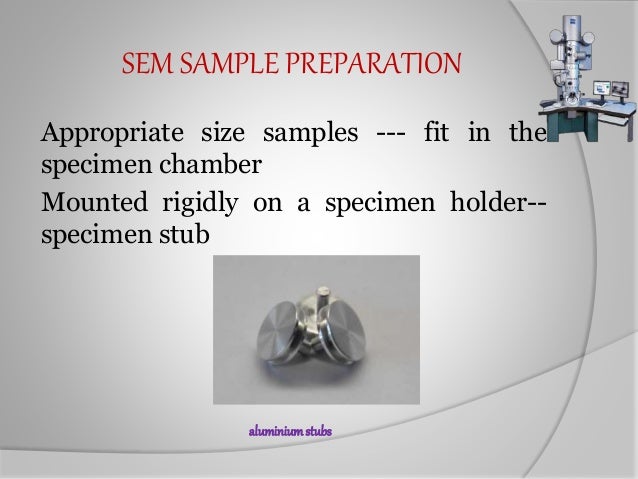
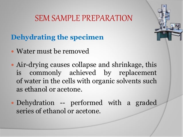
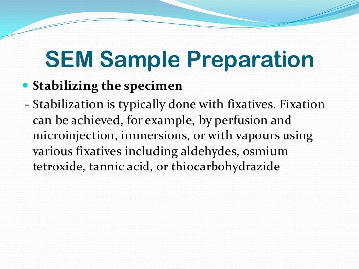
Sample Preparation Scanning Electron Microscope Sem
Scanning Electron Microscope Sem
Scanning Electron Microscope Sem
Scanning Electron Microscope Sem

Cryo Fib Sem Sample Preparation And Integrity Control Cyttron
Scanning Electron Microscopy

Serial Block Face Scanning Electron Microscopy Sem Analysis Workflow Download Scientific Diagram
This video describes how to prepare a sample for use with a scanning electron microscope.
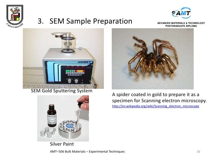
Sample preparation scanning electron microscope sem. They enable scientists to view cells tissues and small organisms in very great detail. The microscope s detection capacity is as much as 1µm from the sample surface. Ideally the smallest representative sample size is the one to use. Sample preparations are essential in scanning electron microscopy.
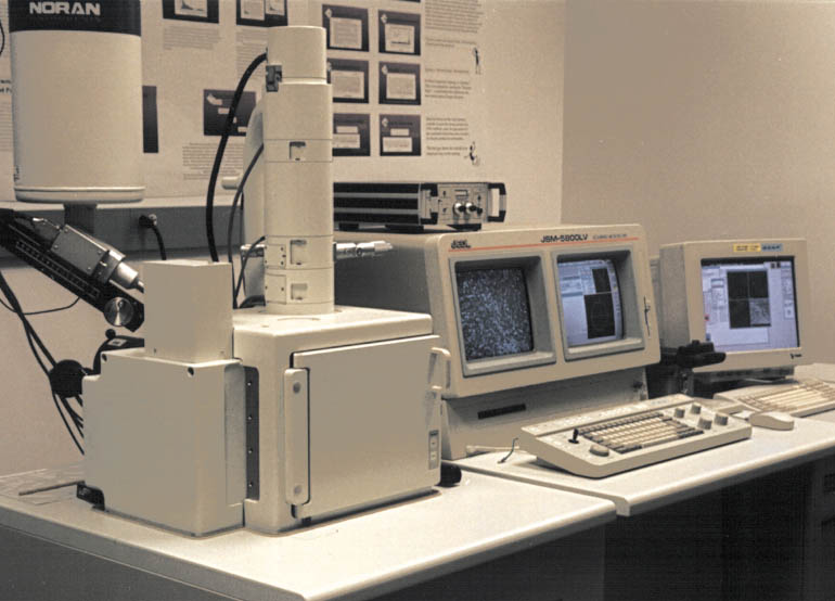
However these biological samples can t be viewed on electron microscopes whilst alive. You need to consider the sample s size shape state and conductive properties prior to sample preparation. The signals that derive from electron sample interactions reveal information about the sample including external morphology texture chemical composition and crystalline structure and orientation of materials making up the sample. Specifically the hatachi tm3000 model of desktop sem.
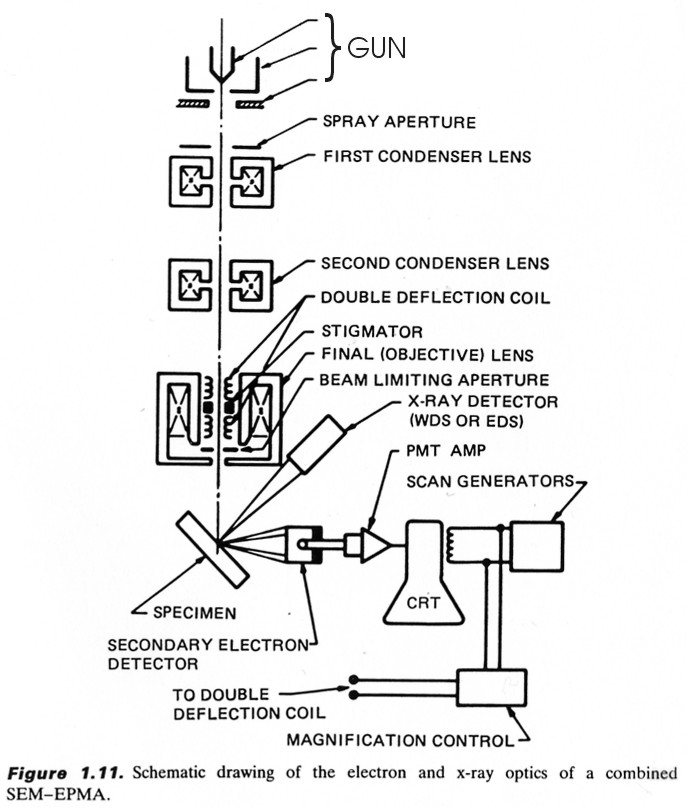
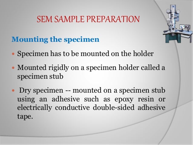
Flawed sample preparations can undermine the quality of results and lead to false conclusions. To learn more about sample preparation for scanning electron microscopes fill out this form to speak with an expert. Plant tissues must be preserved by dehydration for observation in an electron microscope because the coating system and the microscopes operate under high vacuum and most specimens cannot withstand water removal by the vacuum system without distortion 1. The scanning electron microscope sem uses a focused beam of high energy electrons to generate a variety of signals at the surface of solid specimens.
The phenom desktop sem combines superb imaging power up to 100 000x and outstanding technical performance with better depth of focus and chemical contrast. Proper sample preparation plays an important role in obtaining the required information when using scanning electron microscopy sem. Scanning electron microscopy sem is an ideal technique for examining plant surfaces at high resolution. Sample preparation is critical to obtaining high quality sem images.
The electron beam is scanned in a raster scan pattern and the position of. In order to properly interpret images of nerve cells observed in the scanning electron microscope em it is of fundamental importance that fixation dehydration critical point drying cad and metal deposition be accompanied with minimal distortion of nerve and glial cell structures and their cytoarchitectonic arrangement within a gray center. Electron microscopes are very powerful tools for visualising biological samples. The phenom desktop scanning electron microscope sem helps customers stay competitive in a world where critical dimensions are continuously getting smaller.
By taking the time to learn the specific techniques required for different types of samples you can ensure you always obtain the best results.

Cryogenic Specimen Prep Technical Data Sheet
Scanning Electron Microscopy Sem
Scanning Electron Microscopy Sem

Transmission Scanning Electron Microscopes Ppt Video Online Download

Sample Preparation Analytical Testing Laboratory

Sample Preparation Consumables Electron Microscopy Center

A Schematic Of The Strategy For Sample Preparation Scanning Electron Download Scientific Diagram
Experimental Techniques Sem

Preparing Samples For The Electron Microscope Science Learning Hub

Scanning Electron Microscope Definition Images Uses Advantages Facts Britannica
Scanning Electron Microscope Sem

Scanning Electron Microscopes Sem Introduction To Jeol Products Jeol Ltd

Schematic Representation Of The Sample Preparation Procedures In Om Download Scientific Diagram
