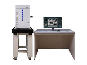
Scanning Electron Microscope Sem
As the intensity of the generated secondary electrons varies depending on the angle of the incident electrons onto the specimen surface subtle variations in the roughness of the surface can be expressed according to the signal intensity.
Scanning electron microscope sem. These images provide information on. Scanning electron microscope advantages and disadvantages in imaging components and applications. The scanning electron microscope sem normally detects secondary electrons to form an image for observation. A scanning electron microscope sem is a type of electron microscope that produces images of a sample by scanning the surface with a focused beam of electrons the electrons interact with atoms in the sample producing various signals that contain information about the surface topography and composition of the sample.
When the electrons strike the specimen a variation of the signal produces an image of the surface or its elemental composition together with. Scanning electron microscopy sem is a powerful materials analysis technique. In fact depending on the sample we dare say the resulting photos often verge on art. The signals that derive from electron sample interactions reveal information about the sample including external morphology texture chemical composition and crystalline structure and orientation of materials making up the sample.
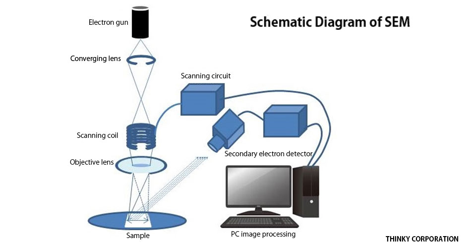
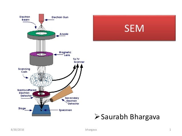
The electron source and electromagnetic lenses that generate and focus the beam are similar to those described for the. Scanning electron microscope sem type of electron microscope designed for directly studying the surfaces of solid objects that utilizes a beam of focused electrons of relatively low energy as an electron probe that is scanned in a regular manner over the specimen. The electron beam is scanned in a raster scan pattern and the position of. The primary electron beam which is produced under high vacuum is scanned across the surface of a specimen.
A scanning electron microscope sem scans a focused electron beam over a surface to create an image. Scanning electron microscopes sem scan a sample with a focused electron beam and get images with information about the samples topography and composition. Scanning electron microscopy sem images the sample surface by scanning it with a high energy beam of electrons in a raster scan pattern. A schematic representation of an sem is shown in figure 1.
A scanning electron microscope sem is a powerful magnification tool that utilizes focused beams of electrons to obtain information. The electrons in the beam interact with the sample producing various signals that can be used to obtain information about the surface topography and composition. The scanning electron microscope sem uses a focused beam of high energy electrons to generate a variety of signals at the surface of solid specimens. Csems conventional sems with a thermic electron source and fe sems field emission sems with a field emission electron source from zeiss deliver high resolution imaging and superior materials contrast.
The high resolution three dimensional images produced by sems provide topographical morphological and compositional information makes them invaluable in a.


