Scanning Electron Microscope Sem With False Coloring

Electron microscope false color scanning electron microscope sem uab university of alabama at birmingham.
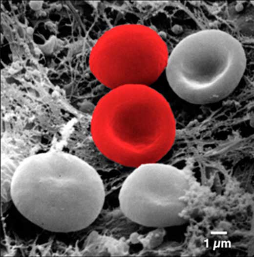
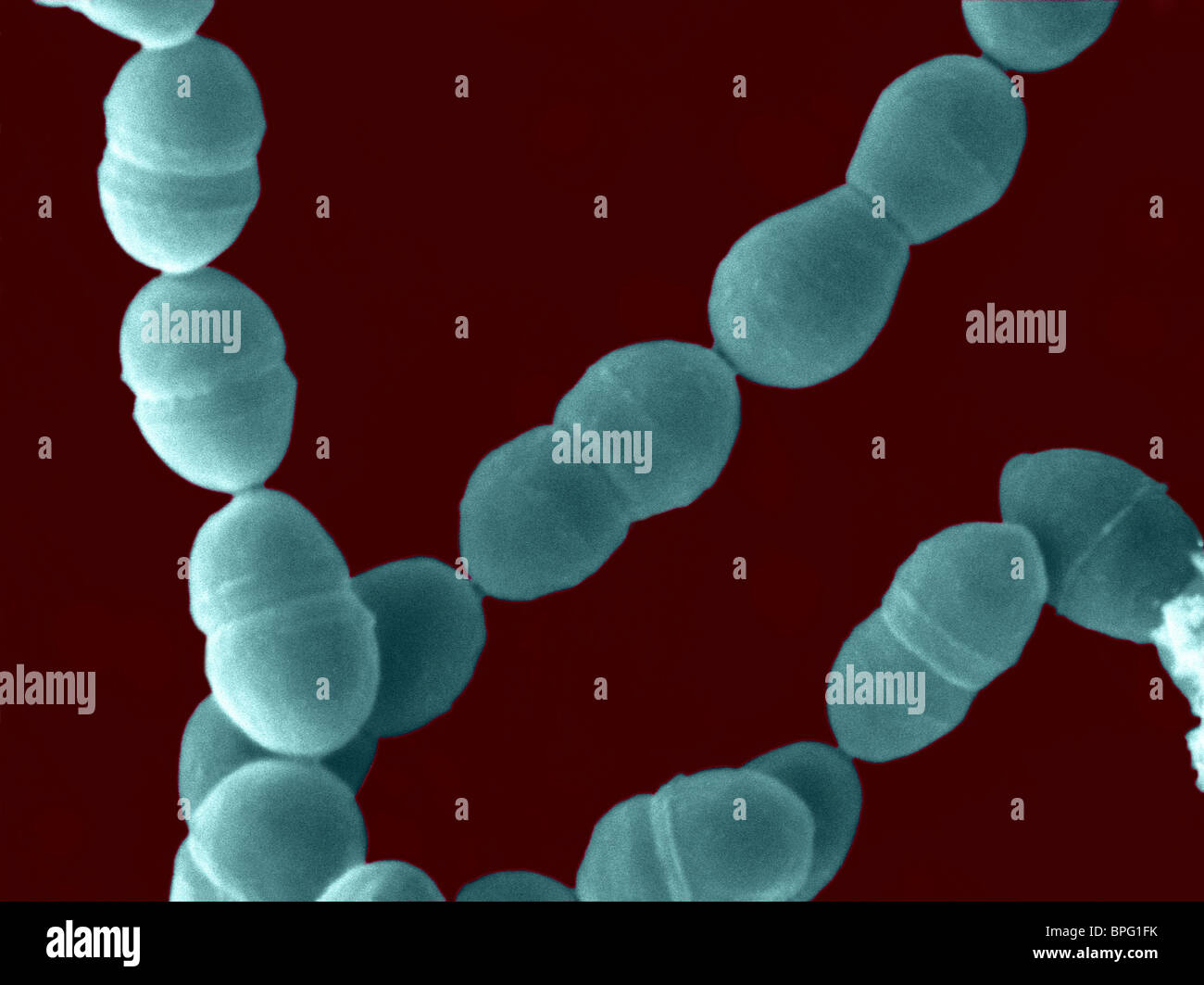
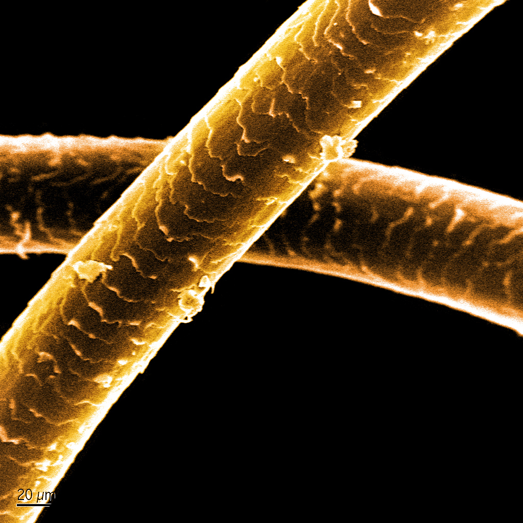
Scanning electron microscope sem with false coloring. The scanning electron microscope sem is widely used in various fields of industry and science because it is one of the most versatile imaging and measurement tools. The electron beam is scanned in a raster scan pattern and the position of. Images produced are particularly appreciated for their high depth of field and excellent image resolution both orders of magnitude better than light microscopy. Nowadays for publishing manuscripts in high impact factor journals sem scanning electron microscope images need to be false colored for enhancing visual illustration.
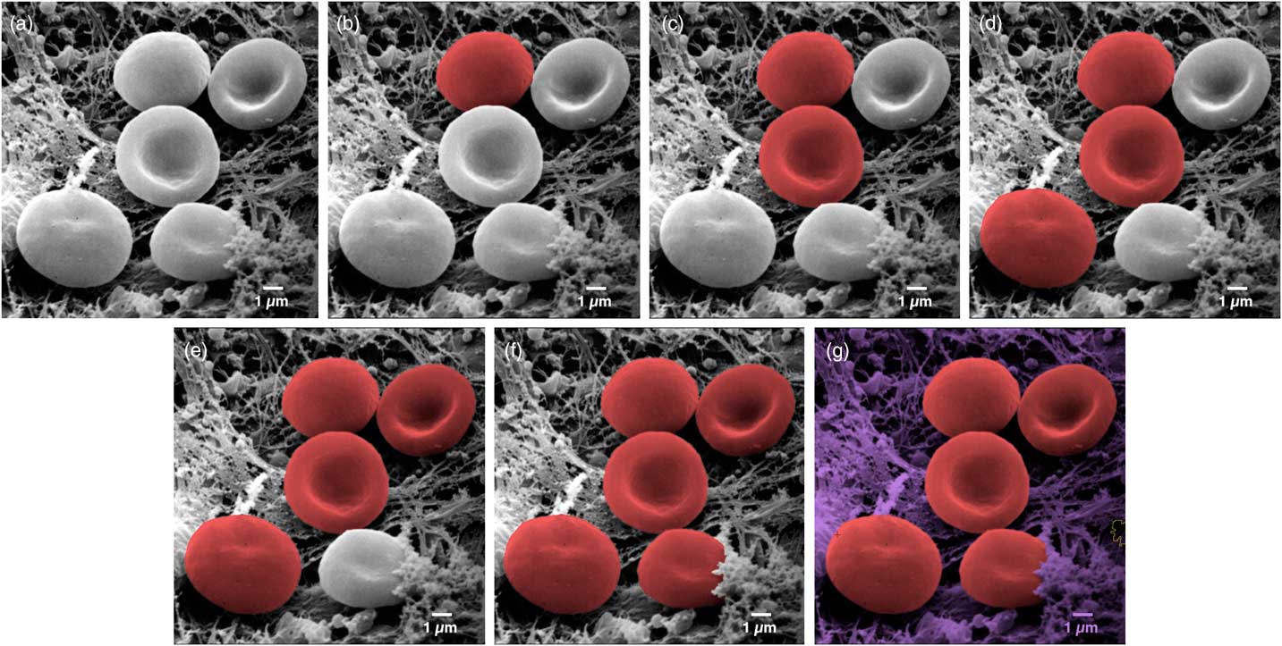
Nowadays for publishing manuscripts in high impact factor journals like science and nature scanning electron microscope sem images need to be false colored for enhancing visual illustration there are several hypothetical ways of adding color to the sem images. A scanning electron microscope sem is a type of electron microscope that produces images of a sample by scanning the surface with a focused beam of electrons the electrons interact with atoms in the sample producing various signals that contain information about the surface topography and composition of the sample. One way to add color is to use photo processing software. Among the myriad of solutions one could either buy expensive software to do the trick or use matlab codes.
A method of adding multiple colors to the sem image using adobe photoshop cs3 is presented in following tutorial. Leave a comment if you you have any questions or would like to schedule time on the sem. April 23rd 2014 how to make false colored sem images. Raw sem scanning electron microscope images induced by electrons are in gray scale because only light carries color information.
However the false monochrome color containing tones of a single.
