Scanning Electron Microscope Sem Images
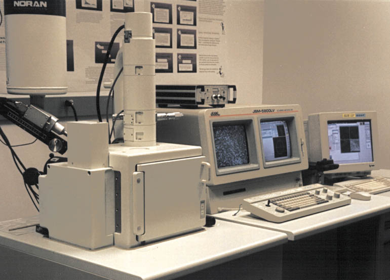
The image is modified and credit goes to wikimedia.
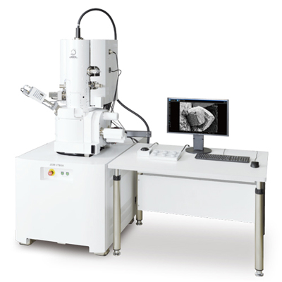
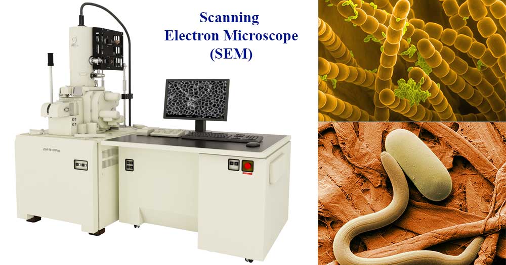
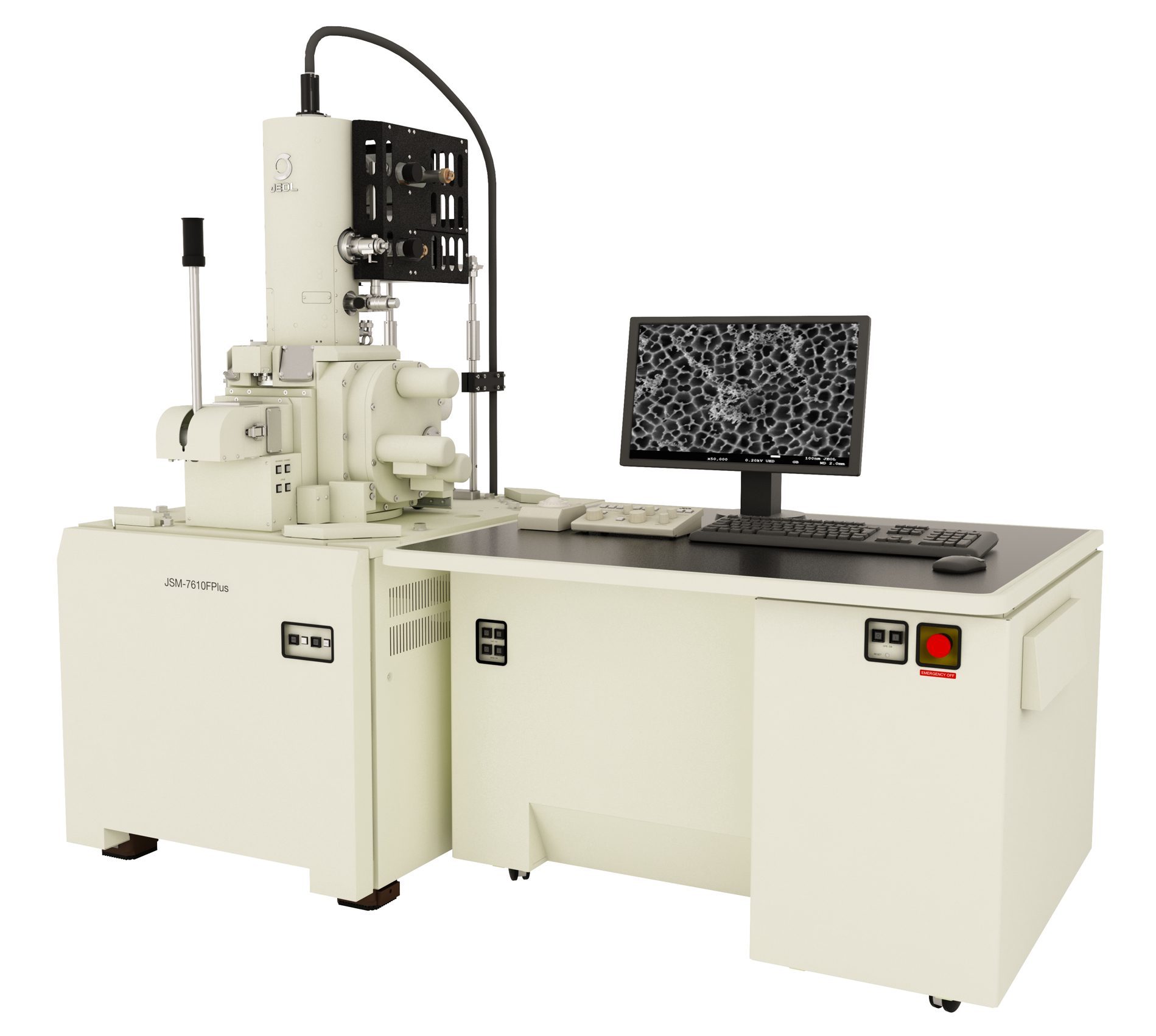
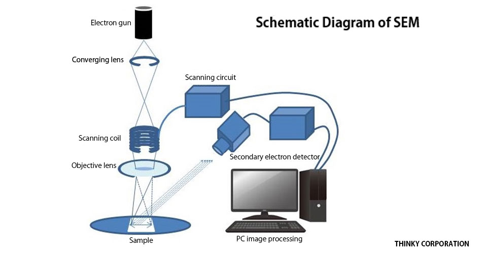
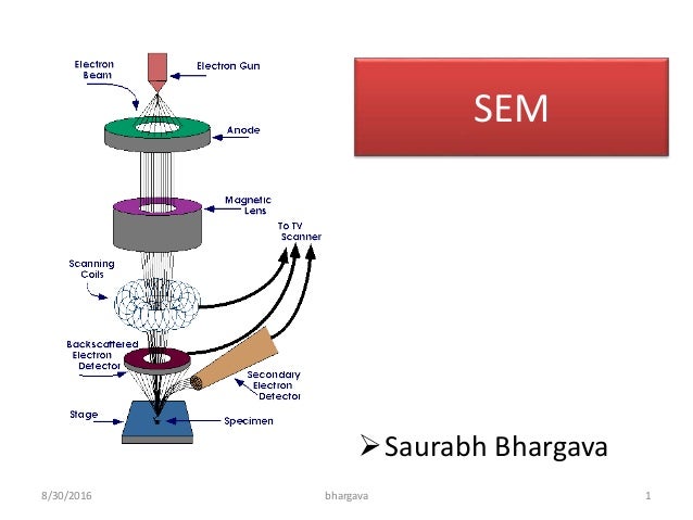
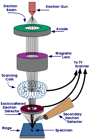
Scanning electron microscope sem images. The scanning electron microscope sem uses a focused beam of high energy electrons to generate a variety of signals at the surface of solid specimens. Power and syred science photo library. X rays or light detector. Electronic gun for the source of the electron.
Scanning electron microscope sem type of electron microscope designed for directly studying the surfaces of solid objects that utilizes a beam of focused electrons of relatively low energy as an electron probe that is scanned in a regular manner over the specimen. Patrick echlin cambridge analytical microscopy cambridge cb4 1xa united kingdom. How to know directly the size using sem. X148 at 8 6 x78 at 10x7cm master size.
The electron source and electromagnetic lenses that generate and focus the beam are similar to those described for the. Component or instrument used in scanning electron microscope. For this sem system a data analysis software is installed. If you want to know the object size from the image there are 2 methods.
Secondary electron detector sed. This paper will consider the ritual that must occur after the final stages of examining the unique se. The process of interpreting images obtained by scanning electron microscopy. Dartmouth electron microscope facility.
A scanning electron microscope sem is a type of electron microscope that produces images of a sample by scanning the surface with a focused beam of electrons the electrons interact with atoms in the sample producing various signals that contain information about the surface topography and composition of the sample. X60 at 6x7cm size. S3400 is made by hitachi. Showing magnified images of materials and objects helps bring context to the world we all live in.
Scanning electron microscopy is an analytical testing method that captures high resolution images of objects as small as 15 nanometers. Coloured scanning electron micrograph sem of superwound guitar string piano wire design. Backscattered electron detector bsed. Scanning electron microscopes are able to achieve a resolution of under 1 nanometer.
The signals that derive from electron sample interactions reveal information about the sample including external morphology texture chemical composition and crystalline structure and orientation of materials making up the sample. Scanning electron microscopy produces images by scanning samples with a focused beam of electrons. The magnification is 1 500 which is displayed at bottom of the image. In this sem image type of sem is displayed.


